PhenoCycler-Fusion
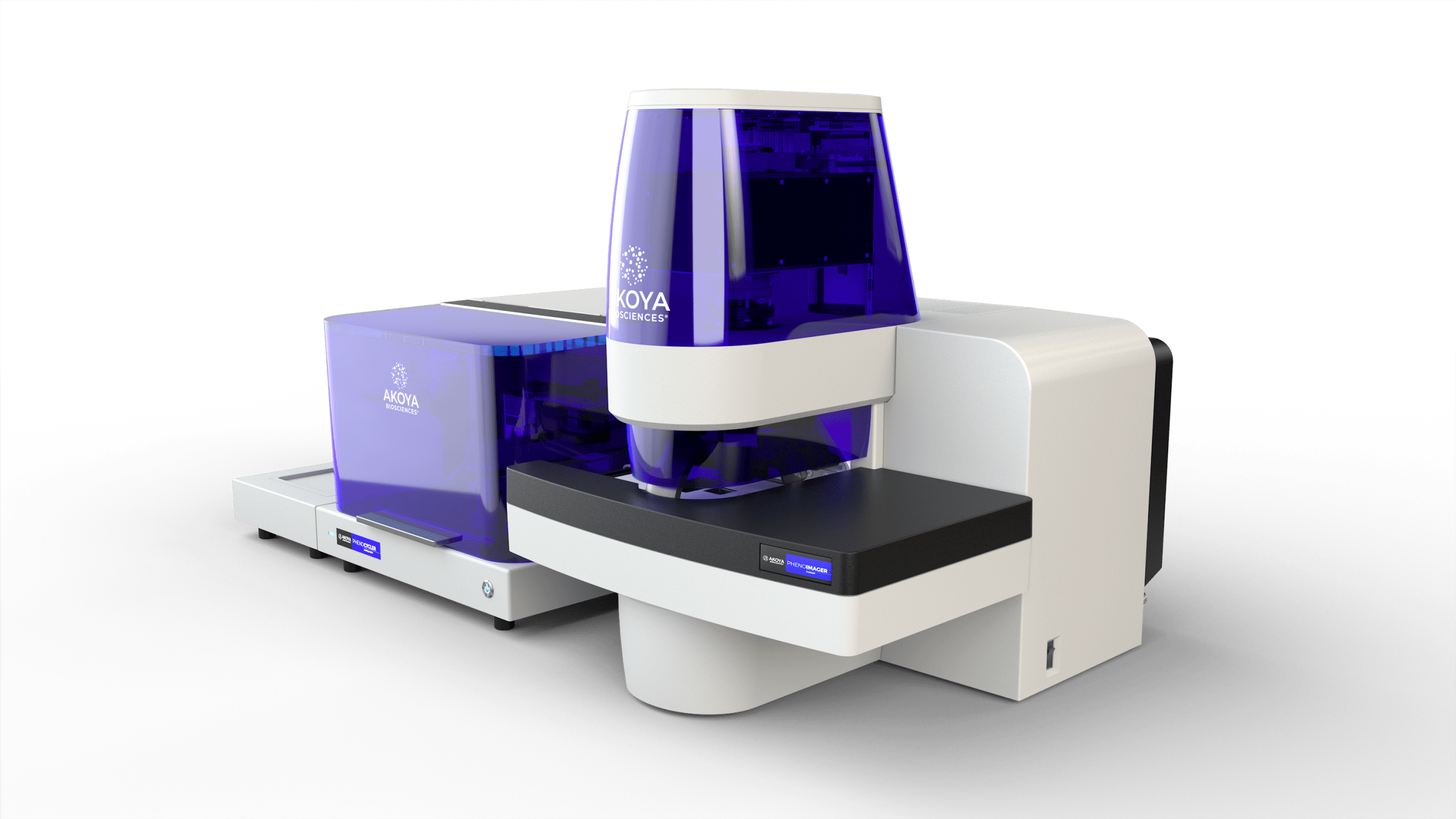
PhenoCycler (PhenoCycler-Fusion) is an imaging-based spatial proteomics platform from Quanterix, formerly Akoya Biosciences; Quanterix completed its acquisition of Akoya in 2025. Protein targets are detected by cyclic immunofluorescence: antibodies carry short DNA barcodes, and each imaging cycle hybridises fluorescent reporter oligonucleotides to a subset of barcodes, images the whole slide, strips the reporters and repeats, so plex accrues across cycles rather than across spectral channels. The unit offers up to 100 protein targets per slide at single-cell resolution. Panels are assembled from the Quanterix pre-conjugated antibody database (354 antibodies) or from custom antibodies conjugated in-house, and pre-made PhenoCode Discovery panels are available. There is no nucleic-acid readout: the DNA barcodes are an addressing mechanism and are never read as sequence, so sequencing is not applicable.
Specification
| Vendor | Quanterix |
|---|---|
| Modality | imaging-based spatial proteomics |
| Targets / plex | up to 100 targets (PhenoCycler-Fusion) |
| Resolution | 20x objective |
| Species | antibody-dependent |
| Billing | slide |
Accreditations & experience
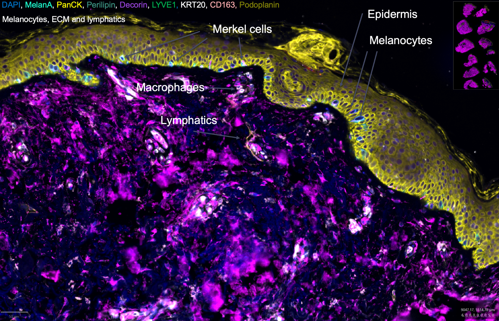
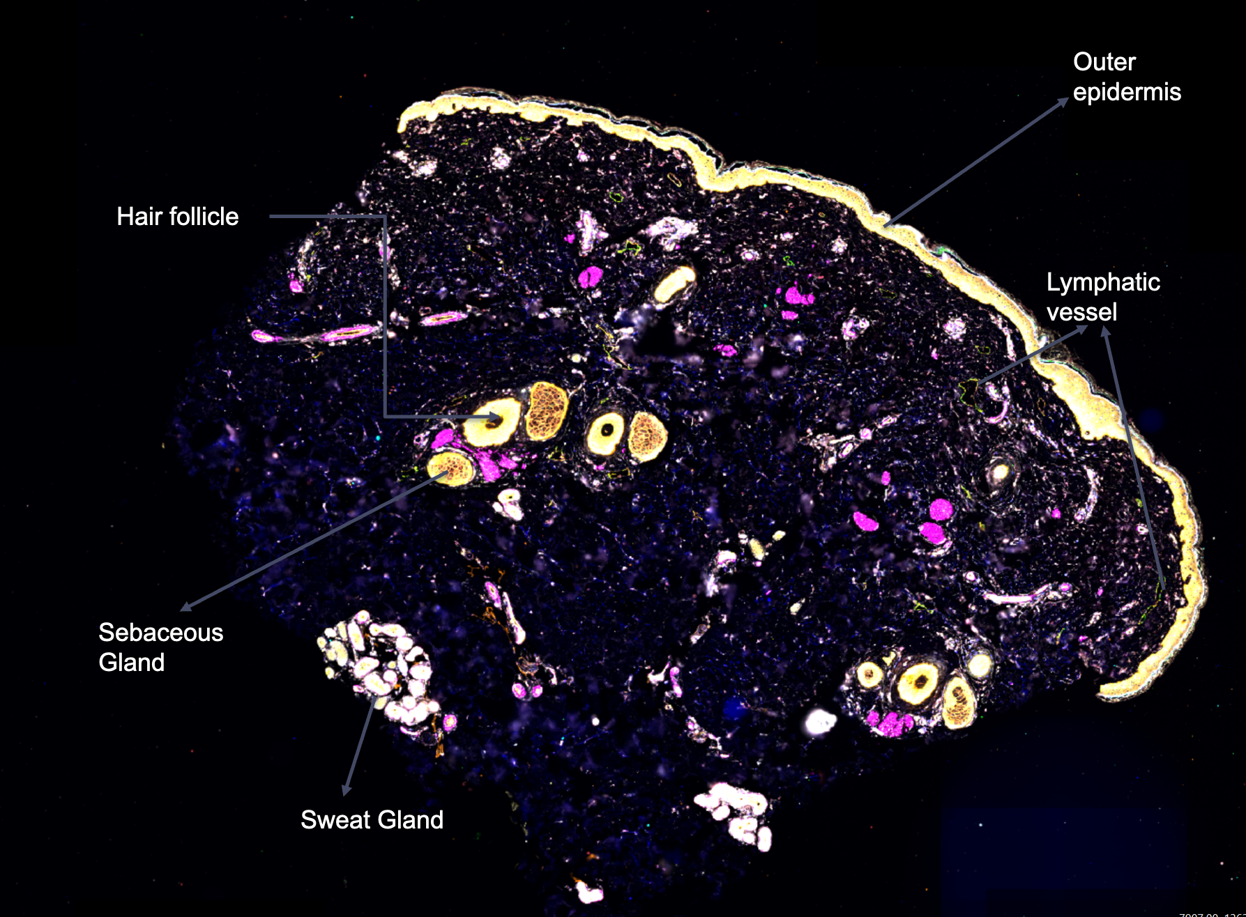
The Spatial Technologies Unit has run extensive custom panels spanning more than 200 protein targets for human and mouse, with the largest panels combining more than 60 antibodies. As an NIH HuBMAP Tissue Mapping Center, the unit has established validated, highly optimized high-plex panels (more than 40 targets) and contributed to the community standards for Antibody Validation Reports (AVRs) and Organ Mapping Antibody Panels (OMAPs).
Panels
PhenoCycler panels are assembled from individual antibodies rather than fixed catalogue modules.
- Pre-conjugated antibodies: Quanterix maintains a database of 354 pre-conjugated antibodies (224 Screened, 104 Inventoried, 26 Community), validated across human, mouse, non-human primate and rat tissue. The full table with target, phenotype, species, tissue preparation and category is held at content/reference/quanterix_phenocycler_antibodies.csv. Inventoried antibodies are pre-conjugated by Quanterix; Screened and Community antibodies require molecular barcoding before use.
- Custom antibodies: any antibody can be used once conjugated with the PhenoCode molecular-barcoding kit. Conjugation is a required step for any antibody that is not already pre-conjugated.
- Pre-made PhenoCode Discovery panels (integrate into the PhenoCycler-Fusion workflow): Human IO60 (60 immuno-oncology markers), Mouse IO (20+ markers), Human Metabolism Spike-In module (extends IO60), Human Neurobiology (42 markers) and Mouse Neurobiology.
Additional capabilities
- Same-section pairing: the unit has run PhenoCycler-Fusion after Xenium on the same tissue section, pairing spatial proteomics with spatial transcriptomics.
- Single staining, no bleaching: PhenoCycler-Fusion stains the sample once, with no antibody bleaching or stripping, so signal quality is maintained from the first antibody to the last while tissue structure is preserved.
- Post-run H&E: same-section H&E can be performed after the run.
- Antibody reference: the full validated-antibody table (target, phenotype, species, tissue preparation, category) is available at the PhenoCycler antibody table.
What you receive
- multiplex whole-slide images (QPTIFF / OME-TIFF)
- cell segmentation and per-cell protein-expression tables
- DAPI morphology and H&E where applicable
- MD5 checksums at transfer
Access and priority
Internal clients (BIDMC, DF/HCC), external academic and non-profit clients, and industry clients all have equal priority. Projects begin upon sample receipt.