About the Spatial Technologies Unit
The Spatial Technologies Unit is a Center of Excellence for the Development and Application of Single Cell and Spatial Tissue Profiling at Beth Israel Deaconess Medical Center and Harvard Medical School.
Spatial Technologies, including Spatial Transcriptomics and Proteomics, were deemed Method of the Year 2020 by Nature Methods and are transforming biomedical research.
Our mission
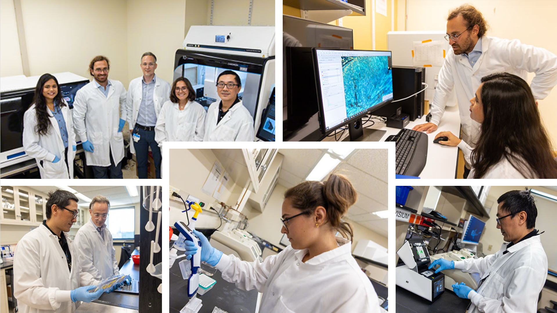
The STU has been established to render cutting-edge spatial and single cell technologies accessible to all, as well as to provide expertise, training, and education, to academia and industry.
By colocalizing spatial technologies with extensive supportive equipment (NGS, tissue processing, imaging, bioinformatics) the STU can provide specialized or A-Z spatial and single cell services, enabling researchers to focus on science and discovery.
The STU bridges study design support, sample processing, advanced instrumentation, and bioinformatic analysis under one roof. It enables rapid innovation, scalability, and translation.
The STU is part of the Harvard Medical School Initiative for RNA Medicine, hosted by the Cancer Center at Beth Israel Deaconess Medical Center.
The unit provides spatial and single-cell services to academia, non-profits, startups, and industry: super-resolution 3D microscopy (Bruker Vutara VXL), full-service single-cell and single-nucleus experiments on the 10x Genomics platform, and a broad array of spatial transcriptomics and proteomics assays. See the technologies for current platforms, specifications, and capabilities, or the equipment page for the full equipment base.
From sample to data
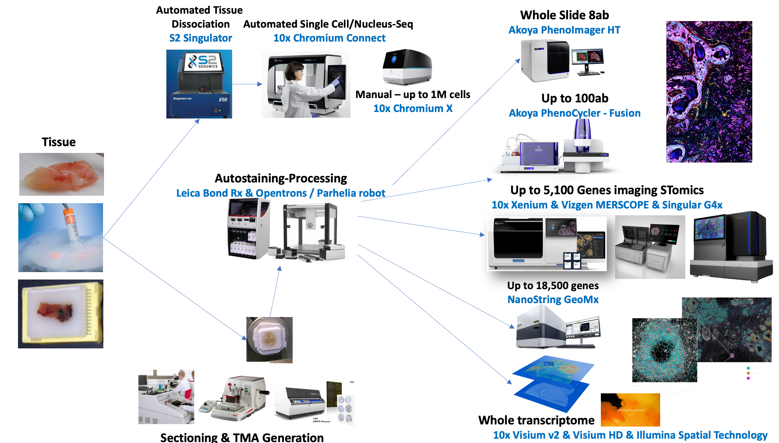
Study design, sample QC, assay, imaging or sequencing, data processing, quality control, transfer, and retention are handled under one roof, so investigators can focus on the science.
Data analysis and bioinformatics
The unit runs the full computational pipeline in house, staffed by dedicated computational biologists. Standard processing follows vendor best practices or STU-optimized pipelines (including more than ten optimized tissue-segmentation pipelines) and delivers processed count matrices, cell segmentation, spatial coordinates, clustering and UMAP embeddings, and post-processing QC in standard formats (AnnData, Seurat, Giotto).
Extended post-processing covers cell filtering and cleanup, batch-effect estimation and harmonization, dimensionality reduction, clustering, and in silico cell-type annotation. Advanced analyses, available as scoped add-ons, include data integration, spatial neighborhood and niche analysis, in situ cell-cell communication, functional analyses, group comparisons, mutation calling and neoantigen discovery, non-coding RNA analysis, and publication-ready figures. The team is among the few worldwide able to integrate single-cell datasets exceeding 300 million cells, and developed the interactive applications for the Human BioMolecular Atlas Program (HuBMAP) Cell-Specific Atlases.
Quality control is extensive, spanning sample receipt through downstream analysis, with more than 500 metrics tracked for a single-cell project and more than 300 for a bulk RNA project. Processing workflows can be containerized (Nextflow) with references, parameters, and run metadata archived per dataset, and the unit routinely formats and deposits data to GEO, dbGaP, HuBMAP, and CELLxGENE-compatible resources.
Clients who want to explore their data without local bioinformatics resources are supported with web applications: the unit's browser-native Spatial Viewer (SViewer) for whole-slide spatial data, and interactive Shiny apps (more than 100 currently hosted) for atlas-level exploration.
Foundation models
Foundation models are revolutionizing how H&E images can be used and integrated with -omic modalities. Our experienced team can help you design novel models, or leverage and optimize cutting-edge solutions, to turn H&E images into a treasure-trove for innovation.
Established and supported by
The unit was established with a capital investment from the Massachusetts Life Sciences Center together with Beth Israel Deaconess Medical Center and the HMS Initiative for RNA Medicine, and is affiliated with the Dana-Farber/Harvard Cancer Center.