GeoMx - Bruker
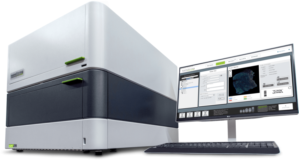
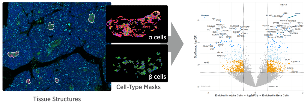
The GeoMx Digital Spatial Profiler (Bruker) is a region-of-interest, morphology-guided spatial profiling platform for formalin-fixed paraffin-embedded (FFPE) and fresh-frozen tissue sections. Tissue is stained with fluorescent morphology markers together with oligonucleotide-barcoded RNA (in situ hybridization) or antibody probes; user-selected regions of interest are illuminated with UV light to release the barcodes, which are read out by Illumina next-generation sequencing and mapped back to each region. RNA and protein are profiled from the same section, separately or simultaneously. The workflow is non-destructive to the section and integrates with a standard histology staining workflow; at the STU, slide staining is automated on the Leica BOND RX. GeoMx is offered through consultation rather than as a fixed-price catalog assay.
Specification
| Vendor | Bruker |
|---|---|
| Modality | region-based spatial profiling, NGS readout |
| Tissue | FFPE, frozen |
| Targets / plex | protein panel 570+ antibodies |
| Species | human and mouse (WTA and CTA panels) |
Accreditations & experience
The unit was among the first to run the GeoMx Whole Transcriptome Atlas (March 2021), work that contributed to one of its earliest spatial tissue-profiling publications (Delorey et al., Nature, 2021). The GeoMx pipeline is automated on the Leica BOND RX, giving the unit close control over throughput and data quality.
Panels
GeoMx profiling is defined by regions of interest rather than a fixed capture area. The unit runs all GeoMx atlases.
- Whole Transcriptome Atlas (WTA): genome-wide RNA profiling (18,000+ genes).
- Cancer Transcriptome Atlas (CTA): a focused panel for cancer biology and the immune response.
- IO Proteome Atlas (IPA): 570+ protein targets for immuno-oncology.
- Custom targets can be added by spike-in (see Additional capabilities).
Multi-omics
RNA and protein are measured on the same FFPE section (same-slide multiomics) or on serial sections. The Whole Transcriptome Atlas or Cancer Transcriptome Atlas can be combined with the 570+ IO Proteome Atlas to profile thousands of transcripts and hundreds of proteins from a single section; updated probe chemistry enables same-slide co-detection at the same sequencing depth and cost as separate detection on serial sections. Up to four fluorescently labeled antibodies or in situ hybridization probes serve as morphology markers to image the tissue and guide region selection.
Additional capabilities
- Region-of-interest geometry: regions can be laid out as geometric shapes to sample heterogeneity, drawn as amorphous shapes that follow tissue-structure boundaries, segmented into two or more compartments by staining pattern (e.g. tumor vs. microenvironment), or placed as concentric contour rings at set distances from a feature or boundary.
- Sample inputs: whole tissue sections, tissue microarrays (TMAs) and organoids.
- Custom targets: up to 400 RNA targets can be spiked into an NGS-readout assay, and up to 40 custom antibodies can be added to an NGS-readout protein assay, including exogenous, endogenous, microbial, viral or non-human targets.
- The workflow is non-destructive to the tissue section and requires no specialized slides.
What you receive
- FASTQ files and raw count matrices
- GeoMx DSP outputs: ROI / segment count matrices
- H&E / morphology images used for region selection
- post-processing QC metrics; MD5 checksums at transfer
Access and priority
Internal clients (BIDMC, DF/HCC), external academic and non-profit clients, and industry clients all have equal priority. Projects begin upon sample receipt.