The 10x Genomics Visium enables a hybridization capture of tissue RNA on slides with 5,000 barcoded spots per area. Slides can have 2 to 4 capture areas of 6.5X6.5mm each. It is a very flexible technology:
- Compatible with both FFPE and fresh frozen tissue samples
- No need to select regions of interest—analyze the whole transcriptome from an entire section.
- 1–10 cell resolution on average per spot depending on tissue type.
- Combine whole transcriptome spatial analysis with immunofluorescence protein detection.
The STU provides end-to-end service, from frozen tissue/paraffin blocks to libraries and/or data. We cover the tissue preparation, optimization, slide loading, visium experiment, library preparation, and sequencing.
For more information about the assay: https://www.10xgenomics.com/products/spatial-gene-expression


10x Genomics Chromium Connect

The 10x Genomics Connect addresses the variability of single cell/nucleus preps by automating the complete process from cell/nucleus suspensions to libraries.
- Generate consistent results: Reduce single cell data variability, generating reproducible and consistent results.
- Characterize single cells by gene expression or immune receptor profiling.
- From sample to sequencing-ready libraries in one day
- Fast turnaround time and maximal consistency to all 10x genomics single cell assays: single nucleus sequencing, single cell sequencing, gene expression + TCR/BCR profiling (VDJ sequencing), and more!
See how the 10x Genomics Connect maximizes single cell robustness and reproducibility https://support.10xgenomics.com/single-cell-gene-expression/index/doc/technical-note-chromium-connect-consistent-automated-single-cell-gene-expression-library-generation
Please follow this link for more information on the instrument: https://www.10xgenomics.com/instruments/chromium-connect
10x Genomics Xenium
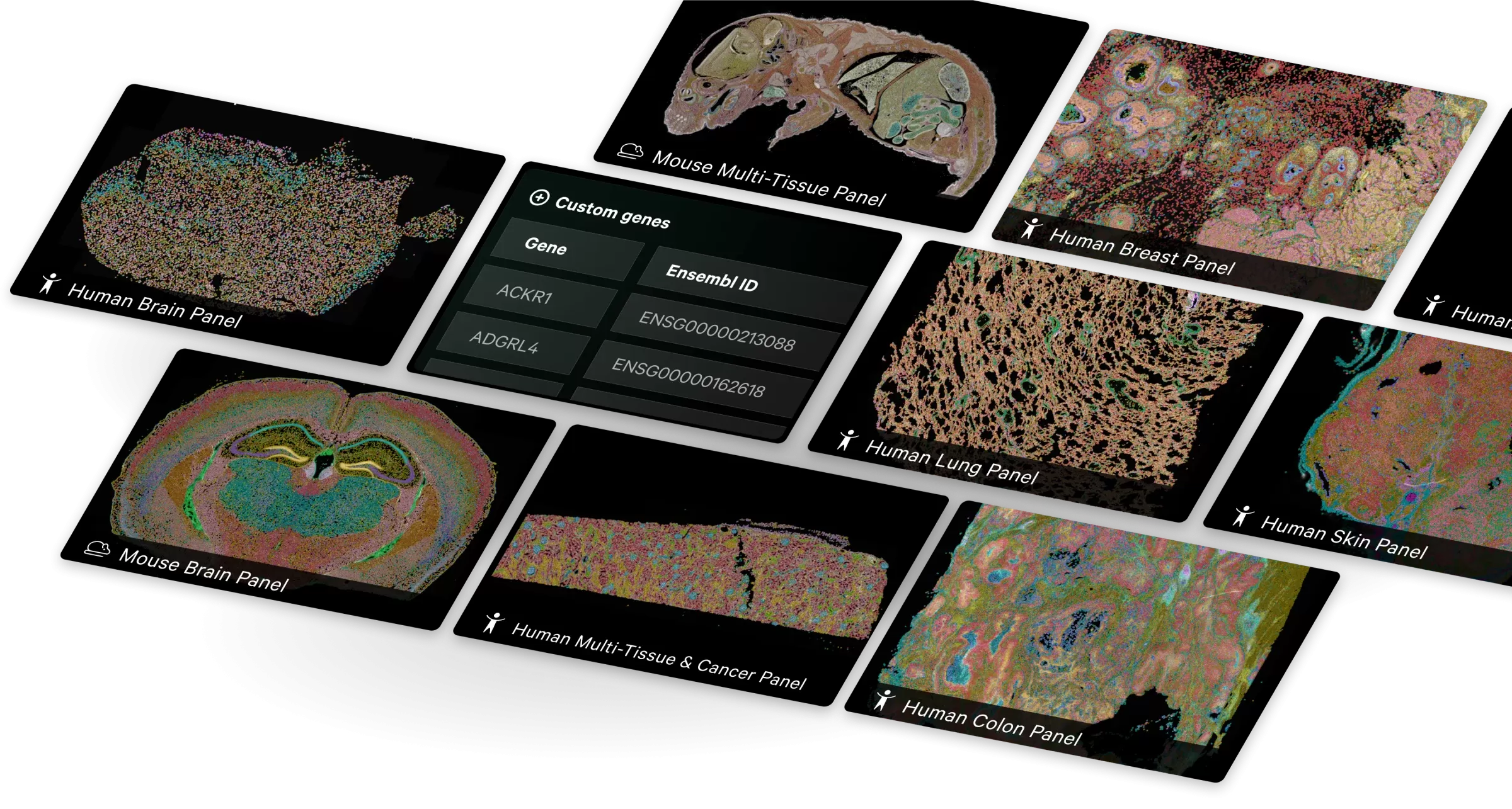
[Place Holder]
Please follow this link for more information on the instrument: https://www.10xgenomics.com/xenium/
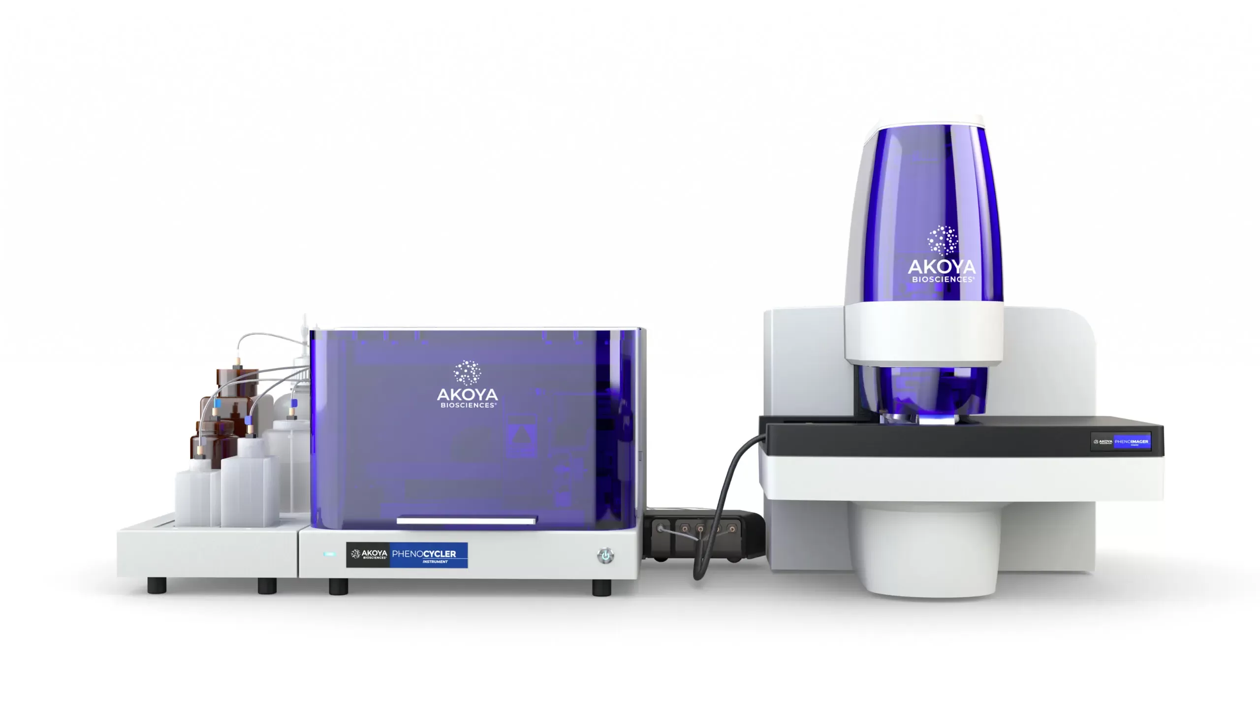
Akoya Polaris Vectra

- State of the art multispectral imaging enables the identification and downstream quantification of multiple overlapping biomarkers (up to 8) without the interference of autofluorescence as the signals are unmixed from one another
- MOTiF™ technology generates unmixed whole slide scans at 40x magnification of up to 7 colors in about 20 minutes (15 x 15 mm region) for analysis of biology across the entire slide in a streamlined workflow and without selection bias.
- High speed, low-cost per slide, enabling screening of whole cohorts in a fraction of the cost and time
- Ability to work on protein level, capturing intracellular and surface protein markers
For more information: https://www.akoyabio.com/phenoptics/mantra-vectra-instruments/vectra-polaris/


The Vutara VXL comprehensive biological workstation for nanoscale biological imaging enables super-resolution single-molecule localization microscopy (SMLM) technology in a streamlined system with compact footprint. The system enables research on DNA, RNA and proteins, from macromolecular complexes and super-structures, to chromatin structure and chromosomal substructures, to studying functional relationships in genomes and in various subcellular organelles. It supports advanced spatial biology research in extracellular matrix structures, extracellular vesicles (EV), virology, neuroscience, and live-cell imaging. It is combined with Bruker’s microscope fluidics unit, Vutara VXL enables multiplexed imaging for targeted, sub-micrometer multiomics in genomics, transcriptomics, and proteomics research.
- Achieves depths of >30 μm imaging with proprietary biplane technology.
- Provides easy yet unlimited multiplexed imaging for spatial genomics, transcriptomics, and proteomics.
- Proprietary biplane technology, combined with a spatial filter in the emission light path, allows you to acquire 3D data with every acquisition. For thicker specimens, the Vutara allows you to easily perform a Z series and automatically localizes and reconstructs the entire volume.
- Making cultured cells, cell colonies, tissue sections, and entire model organisms accessible for single-molecule localization experiments.
- The platform with SRX software is also capable of performing ORCA (Optical Reconstruction of Chromatin Architecture)
Please follow this link for further information https://www.bruker.com/en/products-and-solutions/fluorescence-microscopy/super-resolution-microscopes/vutara-vxl.html


Vizgen MERSCOPE


- Massively multiplexed, error-robust, single-cell in situ transcriptomic imaging
- Quantify and localize RNAs in any tissue sample easily, efficiently, and accurately
- Detect low expressing genes that can be missed using other technologies.
- Localize RNA transcripts at a subcellular level with ≤100 nm resolution.
- High multiplexing with custom gene panel design. Current chemistry up to 500 genes.
- Measure your specified genes on many sample or tissue types.
For more information please visit: https://vizgen.com/products/
Epredia Tissue Microarrayer TMA GrandMaster


- The TMA Grand Master is a fastt, high-capacity and user-friendly tissue microarrayer
- Allows the precise selection and extraction of specific regions of interest on the donor blocks and their insertion in recipient blocks
- It enables us to maximize the utility for the spatial technologies available in the STU and minimize costs by combining multiple regions, blocks, samples, and patients on the same slide. The TMA GrandMaster makes possible the screening of whole patient cohorts in just a handful of slides, minimizing costs and time.
- TMA blocks can be created by combining 0.6, 1, 1.5 and 2 mm diameter tissue cores. The instrument can also extract tissue samples to standard 0.2 ml PCR tubes. Then, the extracted tissue samples can be used later for various applications at the field of molecular pathology.
- 558 at 0.6mm, 286 at 1mm, 135 at 1.5mm, or 84 at 2mm tissue cores can be combined in a single block with unparalleled precision!
For more information please visit: https://www.3dhistech.com/research/tissue-microarrayers/tma-grand-master/
S2 Genomics Singulator 100

The bench-top Singulator System and its single-use cartridges enable reproducible, rapid and hands-off tissue dissociations into single-cell or nuclei suspensions. Its ability to perform cold dissociation minimizes the expression of stress-related genes in cells and helps preserve RNA quality in nuclei. Enables us to easily obtain suspensions of nuclei or high-viability cells for a wide range of single-cell analyses.
Ideal for genomics, cell biology and other ‘omics applications, including scRNA-Seq, snRNA-Seq, ATAC-Seq, CITE-Seq, FACS, and immuno-oncology. S2 Genomics provides a selection of pre-set protocols and pre-formulated reagents for cell isolations for an expanding set of mouse, rat, and human tissues, including tumors.
Please visit here for more information: https://s2genomics.com/singulator-100/